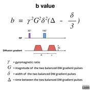
mri b value
To explore the optimal b value in diffusion-weighted imaging DWI for differentiation of benign and malignant abdominal lesions. The b value is used in MRI in the context of Diffusion Weighted Imaging DWI.
Diffusion Weighted Imaging Radiology Reference Article Radiopaedia Org
DW images of eight healthy volunteers were obtained using.

. Crossref Medline Google Scholar. Two recent studies explored DWI at an ultra high b-value of 2000smm 2. DICOM enhanced MRI b-value field 00189087 has been populated in all images with the appropriate b-value.
Required if Frame Type. To test new models eg. Constant b in GutenbergRichter law.
BACKGROUND AND PURPOSE. Image order was standardized. DWI is done to determine the rate of molecular diffusion in different areas of the body.
A low threshold b0. In general in healthy. Answer 1 of 2.
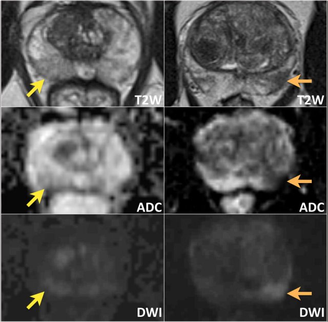
High b-value diffusion-weighted imaging DWI provides different features not appreciated at lower b-value and have been recently studied in several clinical issues. A total of 108 consecutive patients age 60. Diffusion-weighted imaging DWI is commonly used to distinguish between benign and malignant liver lesions.
B 500 smm 2. Important characteristic of a thermistor B parameter B-factor in crystallography. B-value may refer to.
Depending on the organ being imaged b-values typically range from 50-1000smm 2. This is the actual b-value for original frames and those derived from frames with the same b-value or the most representative b-value when derived from images with different b-values. Pretreatment prediction of brain tumors response to radiation therapy using high b-value diffusion-weighted MRI.
In the abdomen lower b values applied often range from 50 to 150. The b-value is a factor of diffusion weighted sequences. Recent technological advances in MR instrumentation allow acquisition of whole-brain diffusion-weighted MR scans to be obtained with b values greater.
Acquisition parameter in diffusion MRI. These can either be calculated directly from the isotropic DWI. Topics referred to by the.
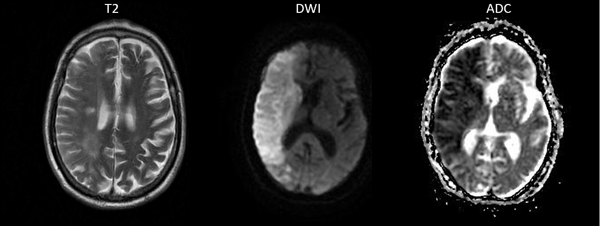
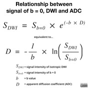
The ADC map in contrast is related to the natural logarithm ln of the isotropic DWI divided by the initial T2 signal b0. Göttingen Mini Pig Brain. A b factor of zero b 0 smm 2 indicates no diffusion weighting and the image is analogous to a T2-weighted image.
To sense slow moving water molecules and smaller diffusion distances b values should be higher eg. However different b-values are recommended. The calculated high b-value DW MRI was then computed by extending this linear equation to the corresponding b- values using the estimated perfusion and diffusion components.
We performed a meta-analysis to assess the diagnostic accuracy of high b-value diffusion-weighted imaging for patients with prostate cancerA comprehensive literature search of the. The higher the value b the stronger the diffusion. All images were acquired using a 15-T clinical MRI system Ingenia Philips Healthcare with a head-array surface coil.
With enhanced gradients the whole b ra in can b e scanned with in seconds. The b factor summarizes the influence of the gradients on the diffusion weighted images. Apparent diffusion coefficient is calculated using different b-values.
The degree of diffusion weight in g correlates with the strength of the diffusion gradients characterized b y the b - value which. Various fibre reconstruction methods high-quality diffusion MRI data sets acquired with multiple b-values is a must. 4-b value ADC maps.
2015 designed a study to correlate.
2
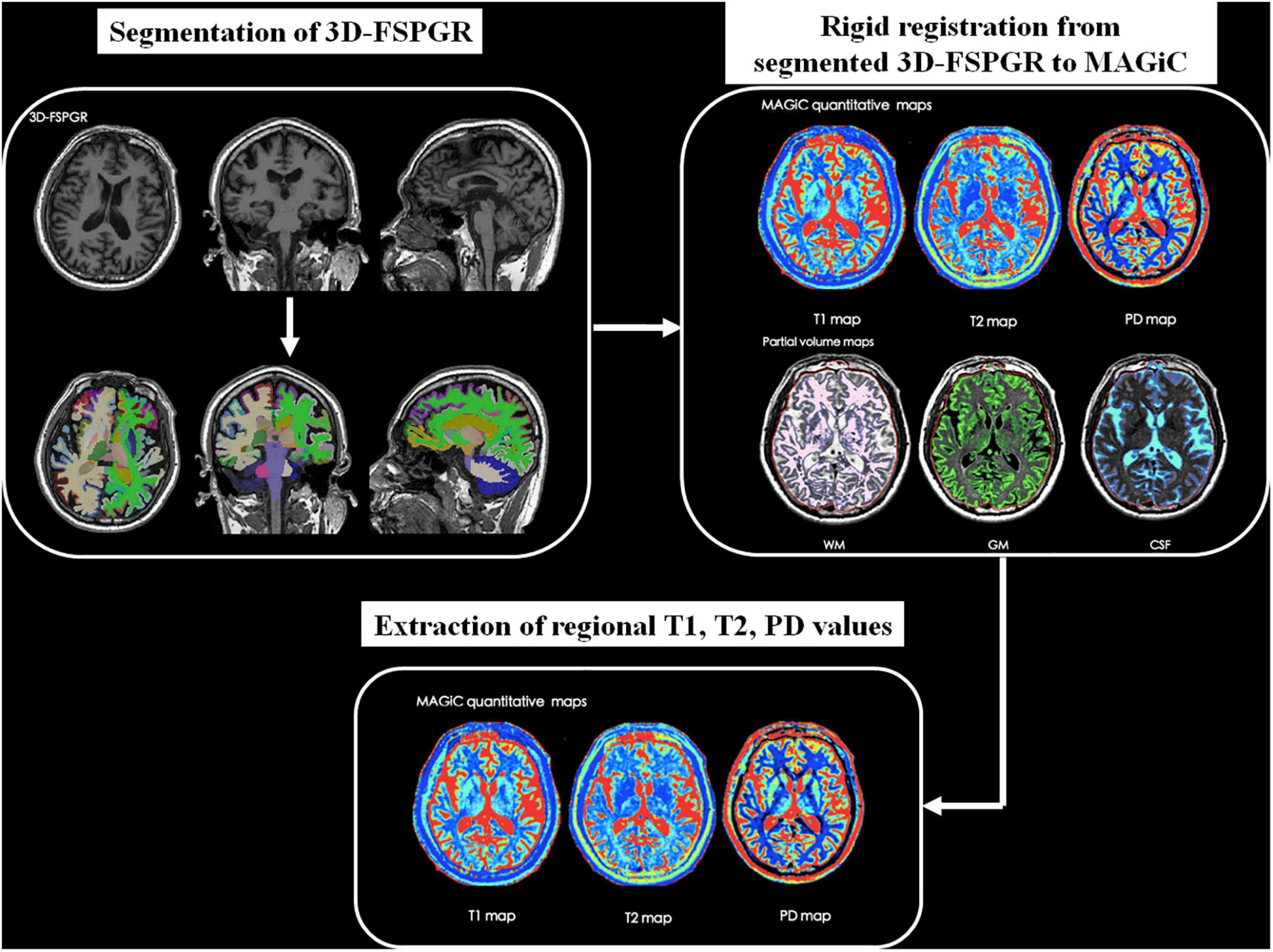
Frontiers Quantitative Analysis Of Synthetic Magnetic Resonance Imaging In Alzheimer S Disease
Apparent Diffusion Coefficient Radiology Reference Article Radiopaedia Org
Diffusion Weighted Imaging In Hemorrhage Radiology Key
2

Magnetic Resonance Imaging Of Brain An Overview Sciencedirect Topics

Principles Of Diffusion Tensor Imaging And Its Applications To Basic Neuroscience Research Neuron
Tensor Valued Diffusion Encoding For Diffusional Variance Decomposition Divide Technical Feasibility In Clinical Mri Systems Plos One

Signal Intensity Of Dwi And Adc In Diffusion Restriction Increased Download Scientific Diagram
The Basics Of Mri Interpretation Radiology Geeky Medics
Diffusion Weighted Imaging Radiology Reference Article Radiopaedia Org

Diffusion Tensor Imaging Dti Fiber Tracking Imagilys
Diffusion Weighted Imaging Radiology Reference Article Radiopaedia Org

Apparent Diffusion Coefficient Radiology Reference Article Radiopaedia Org

Pulse Sequences For Diffusion Weighted Mri Sciencedirect

Apparent Diffusion Coefficient Radiology Reference Article Radiopaedia Org
The Radiology Assistant Prostate Cancer Pi Rads V2
Apparent Diffusion Coefficient Radiology Reference Article Radiopaedia Org

Diffusion Weighted Imaging Radiology Reference Article Radiopaedia Org


Comments
Post a Comment